
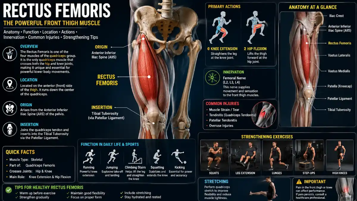
The rectus femoris is one of four quadriceps muscles, and it’s the only one that crosses both the hip and the knee joint. This dual-joint design is what makes it central to kicking, sprinting, and climbing stairs-and also why it’s one of the more injury-prone muscles in field-sport athletes.
This guide covers the muscle’s anatomy, biomechanics, strain grading, diagnosis, and an evidence-informed rehabilitation framework, with clear guidance on when a strain needs medical evaluation rather than self-treatment.
Rectus Femoris Anatomy at a Glance
| Feature | Detail |
| Location | Anterior (front) compartment of the thigh |
| Muscle group | Quadriceps femoris |
| Origin | Direct head: anterior inferior iliac spine (AIIS); indirect head: groove above the acetabulum |
| Insertion | Base of the patella via the quadriceps tendon; force transmits to the tibial tuberosity via the patellar ligament |
| Innervation | Femoral nerve (L2–L4) |
| Main arterial supply | Lateral (circumflex) femoral artery, a branch of the femoral/profunda femoris system |
| Joints crossed | The hip and knee are unique among the four quadriceps muscles |
| Main actions | Hip flexion and knee extension |
What Is the Rectus Femoris?
The rectus femoris is a fusiform, bipennate muscle running down the front of the thigh, and it’s the most superficial of the four quadriceps muscles. It lies on top of the vastus intermedius and overlaps the medial edges of the vastus medialis and vastus lateralis.
Unlike the other three quad muscles, it originates from the pelvis rather than the femur, a structural difference that explains both its unique two-joint action and its comparatively higher injury risk during sport.
- Read more: Rectus Femoris
Origin and Insertion
The muscle has two proximal heads that converge into a common tendon:
- Direct (straight) head-anterior inferior iliac spine (AIIS)
- Indirect (reflected) head-a groove just above the rim of the acetabulum, extending onto the hip joint capsule
These fuse and descend as the rectus femoris, joining the quadriceps tendon to insert at the base of the patella, with force continuing to the tibial tuberosity via the patellar ligament.
Innervation and Blood Supply
The rectus femoris is innervated by the femoral nerve, with root contributions from L2 through L4. Its main arterial supply comes from the lateral circumflex femoral artery, a branch of the femoral and deep femoral (profunda femoris) system, with minor contributions from the superficial circumflex iliac artery.
Rectus Femoris vs. Other Quadriceps Muscles
The rectus femoris is the only quadriceps muscle that acts on the hip; the other three act on the knee alone. This is the single most clinically important distinction within the quad group, and it directly explains the muscle’s unique injury pattern.
| Muscle | Crosses hip? | Crosses knee? | Hip flexion | Knee extension |
| Rectus femoris | Yes | Yes | Yes | Yes |
| Vastus lateralis | No | Yes | No | Yes |
| Vastus medialis | No | Yes | No | Yes |
| Vastus intermedius | No | Yes | No | Yes |
How the Rectus Femoris Moves the Body
The rectus femoris actively produces hip flexion and knee extension; it does not actively produce hip extension or knee flexion. This distinction matters because the muscle is frequently and incorrectly described as generating the opposite motions during a kick.
- Concentric action (what it actively does): flexes the hip and extends the knee, working alongside the iliopsoas at the hip and the other three vastus muscles at the knee.
- Eccentric loading (what stretches and stresses it): the muscle lengthens across both joints simultaneously when the hip extends while the knee flexes at the same time, precisely the position reached during the backswing of a kick or the late swing phase of sprinting. This combined two-joint stretch, followed by a rapid, forceful concentric contraction, is the primary mechanical setup behind most rectus femoris strains.
This behavior is why the muscle is sometimes informally called the “kicking muscle”-not because it produces the kicking motion by itself, but because it’s subjected to unusually high mechanical demand during that exact sequence of movement.
Where Rectus Femoris Pain Is Felt
Symptom location often points to which part of the muscle-tendon unit is affected:
- Proximal, anterior hip pain suggests injury at the AIIS origin, the conjoined tendon, or the proximal myotendinous junction-the most common injury site, particularly at the indirect head.
- Muscle-belly, anterior thigh pain suggests a contusion or myofascial-level strain within the body of the muscle.
- Distal pain, just above the knee, suggests a distal myotendinous junction strain, which is comparatively rare.
Rectus Femoris Strain: Causes and Grading
A rectus femoris strain is a partial or complete tear of the muscle-tendon unit, most often caused by a forceful eccentric contraction, such as kicking a ball or sprinting at high speed.
Common Mechanisms
- Forceful kicking, with soccer being the most frequently reported context
- Sprinting or explosive acceleration
- Jumping and landing, particularly from a height
- Sudden change of direction under load
Strain Grades
Severity is generally classified into three grades, and the grade affects both prognosis and the pace of rehabilitation.
| Grade | Tissue Damage | Typical Signs | Approximate Recovery* |
| Grade 1 | Muscle edema without fiber disruption | Mild pain or tightness, little to no strength loss, no palpable defect | Roughly 1–2 weeks |
| Grade 2 | Partial tear of the myotendinous junction | Sudden sharp pain, swelling, bruising, moderate strength loss, possible small palpable defect | Roughly 4–6 weeks |
| Grade 3 | Complete tear, with or without retraction | Severe pain, marked swelling or bruising, major or complete strength loss, palpable deformity | Roughly 9–12 weeks |
*Recovery ranges are approximate and vary by individual, injury location, and treatment approach; only a clinician can confirm the grade and a realistic timeline. In adolescent athletes, the same kicking or sprinting mechanism can cause an avulsion fracture at the AIIS rather than a muscle tear, because the growth plate there is still developing and structurally weaker than mature bone.
How Is a Rectus Femoris Injury Diagnosed?
Diagnosis typically starts with a clinical examination and is confirmed with imaging when the severity or exact injury location is unclear. A clinician will usually assess pain during resisted hip flexion, resisted knee extension, and passive stretch of the muscle, along with palpation for tenderness, swelling, or a defect.
- Ultrasound is often used as a first-line imaging tool for soft-tissue strains because it’s accessible and cost-effective.
- MRI provides the most detailed picture for grading strain severity and pinpointing whether the direct head, indirect head, or myotendinous junction is involved.
- X-ray is particularly relevant in adolescents or young athletes with sudden proximal hip pain after kicking or sprinting, to rule out an AIIS avulsion fracture.
Differential Diagnosis
Several other conditions can mimic a rectus femoris strain, and distinguishing between them changes the treatment approach:
- Iliopsoas strain-pain sits deeper and more medial in the groin
- Sartorius strain-pain follows the inner thigh line rather than the center of the front thigh
- Quadriceps tendon injury-pain concentrates just above the patella
- Hip joint pathology, such as a labral tear, often presents with clicking, catching, or deep hip pain rather than a discrete muscular tear.
- AIIS is avulsion-specific to adolescents and confirmed on X-ray
When to Seek Medical Care
Seek prompt medical assessment rather than self-treating if any of the following are present:
- Inability to walk normally or bear weight
- Significant swelling or visible bruising
- A palpable gap or defect in the muscle
- Severe weakness or inability to actively extend the knee against resistance
- Sudden proximal hip or thigh pain in an adolescent athlete after kicking or sprinting, which may indicate an avulsion
- Symptoms that persist beyond a few days or worsen despite rest
What to Do After a Suspected Strain
Initial self-care focuses on protecting the injured muscle and avoiding further damage, not on aggressive treatment. This approach is appropriate only for mild symptoms without any of the red flags above.
- Stop the aggravating activity immediately-continuing to kick, sprint, or load the leg risks converting a partial tear into a more severe one.
- Protect the area by avoiding deep stretching or heavy loading in the first few days.
- Move within a pain-free range-gentle, symptom-guided movement is generally preferred over complete immobilization, but should never push into sharp pain.
- Manage swelling with rest, ice, and elevation as needed for comfort.
- Discuss pain relief with a clinician or pharmacist rather than defaulting to anti-inflammatory medication, since appropriateness and dosing depend on individual health factors.
Anyone showing red-flag signs should be evaluated by a doctor or physiotherapist before resuming activity. Clinical grading, sometimes supported by imaging, is usually what determines a safe rehabilitation timeline.
Rehabilitation Progression
Rehabilitation is typically progressive, moving from pain control toward full sport-specific loading rather than jumping straight into strengthening exercises. A general framework-always adjusted to individual grade and clinician guidance-looks like this:
- Pain control and protected motion, gentle range-of-motion work within pain-free limits.
- Isometric loading-static muscle contractions, such as gentle quad sets, once acute pain has settled.
- Progressive resistance-controlled dynamic exercises with light resistance, increasing gradually.
- Eccentric loading-exercises that lengthen the muscle under control, since rectus femoris injuries are eccentric in nature, and eccentric strength helps protect against re-injury.
- Running and kicking drills-sport-specific movement patterns reintroduced at low intensity.
- Return to sport, full-speed, unrestricted training and competition.
Rehabilitation Exercises by Phase
| Exercise | Phase | Goal | Typical Sets/Reps | Stop or Modify If |
| Isometric quad set | Early | Muscle activation without joint movement | 3–5 x 10-second holds | Sharp pain, not mild discomfort |
| Straight leg raise | Early strengthening | Isolated hip flexor and quad activation | 2–3 x 10–15 reps | Pain during the lift or at the end range |
| Seated band leg extension | Progressive resistance | Loaded knee extension strength | 2–3 x 10–12 reps | Pain that lingers after the set |
| Controlled lunge | Functional strengthening | Combined hip and knee loading | 2–3 x 8–10 reps per side | Instability or sharp anterior thigh pain |
| Eccentric knee extension | Eccentric loading | Build tolerance to lengthening stress | 2–3 x 8 reps, slow tempo | Any reproduction of the original injury pain |
Foam rolling and self-massage can support tissue mobility and comfort during rehabilitation, but they’re a mobility tool rather than a strengthening exercise, and shouldn’t replace progressive loading work.
Return-to-Sport Criteria
Rather than relying on a fixed calendar, most rehabilitation approaches use functional benchmarks before clearing an athlete for full sport:
- Pain-free walking and daily activity
- Pain-free resisted hip flexion and knee extension
- Full, symmetrical range of motion compared with the uninjured leg
- Near-symmetrical strength on formal testing
- Sport-specific running and kicking performed without symptoms
Stretching the Muscle Safely
Because this muscle crosses both the hip and the knee, an effective stretch combines hip extension with knee flexion at the same time.
Standing quad stretch: Stand holding a wall for balance, bend one knee, bringing the heel toward the buttock, grasp the ankle, and gently pull back while pushing the hips forward. Hold for 20–30 seconds per side.
Kneeling hip flexor stretch: From a half-kneeling lunge position, tilt the pelvis backward, tighten the glute on the rear leg, and push the hips gently forward. Hold for 30 seconds.
Some clinicians note that a chronically tight rectus femoris often responds better to a combination of targeted strengthening and mobility work rather than stretching alone. Persistent tightness that doesn’t improve with stretching is a reasonable reason to consult a physical therapist.
Key Takeaways
This muscle is anatomically distinct from the rest of the quadriceps because it’s the only one crossing both the hip and knee, giving it a combined role in hip flexion and knee extension. That same feature makes it vulnerable to eccentric-overload strains during kicking and sprinting, which is why accurate grading, appropriate diagnosis, and a structured rehabilitation progression matter more than generic stretching or strengthening advice alone.
Frequently Asked Questions
What is the difference between the rectus femoris and the rest of the quadriceps?
It’s the only one of the four quadriceps muscles that crosses both the hip and knee joint; the vastus medialis, lateralis, and intermedius attach only to the femur and act solely on the knee. This gives it a combined role in hip flexion and knee extension that the other three muscles don’t share.
Why is it sometimes called the “kicking muscle”?
It earns this nickname because kicking places it under unusually high mechanical demand: the backswing lengthens it across both the hip and knee simultaneously, then the forward kick requires a rapid, forceful concentric contraction. This same movement pattern explains why kicking sports carry an elevated risk of injury to this muscle.
Can tightness here contribute to lower back discomfort?
Tightness can pull the pelvis into anterior tilt because of its attachment at the AIIS, and altered pelvic positioning is sometimes discussed as a contributing factor in mechanical lower-back discomfort. This relationship isn’t universal or independently sufficient to cause back pain, so it should be considered alongside core and glute strength rather than as a standalone explanation.
How long does a strain take to heal?
Recovery depends heavily on grade: mild (Grade 1) strains often improve in roughly 1–2 weeks, moderate (Grade 2) strains in roughly 4–6 weeks, and severe (Grade 3) tears in roughly 9–12 weeks, though these are general ranges rather than guarantees. Healing, being pain-free, returning to training, and returning to full competition are different milestones, and return to sport should be based on functional criteria confirmed by a clinician rather than a fixed date.
Is it safe to stretch a strained muscle right away?
No-aggressive stretching immediately after a strain can worsen the tear, so initial care should focus on protecting the muscle and moving within a pain-free range rather than deep stretching. Gradual, controlled stretching is typically reintroduced only once acute pain and swelling have settled, guided by the injury’s grade.
What sports carry the highest injury risk?
Soccer is most frequently associated with strains here due to repeated forceful kicking, and sprint-based sports, along with activities involving explosive jumping or landing, also carry elevated risk. Track and field athletes, particularly sprinters and kickers, are commonly affected.
Why do adolescents sometimes get a bone avulsion instead of a muscle tear?
In younger athletes, the growth plate at the AIIS is still developing and is structurally weaker than mature bone, so a forceful contraction can pull off a small bone fragment rather than tearing the muscle tissue. This is confirmed with an X-ray and is a key reason adolescents with sudden proximal hip pain after kicking or sprinting need medical evaluation.
How is this injury distinguished from an iliopsoas or sartorius strain?
Location and movement testing help differentiate them: iliopsoas strains tend to cause deeper, more medial groin pain, sartorius injuries cause pain along the inner thigh line, while this injury localizes to the front of the hip or thigh and worsens with resisted knee extension or hip flexion. Clinical examination, and imaging when needed, reliably confirms which structure is involved.
What muscles work opposite this one?
The hamstrings are the functional antagonist at both the hip and knee-they extend the hip and flex the knee, the reverse of this muscle’s actions. Balanced quadriceps-to-hamstring strength is considered protective against strain injuries in sprinting and kicking athletes.
Can foam rolling help during recovery?
Foam rolling can support short-term tissue mobility and comfort and is commonly used alongside active movement in rehabilitation, but it’s a mobility tool rather than a strengthening method and doesn’t replace progressive loading exercises. It’s most useful as one part of a broader rehabilitation or maintenance routine, not as a standalone treatment.